Featured


This is a print of the skull and sclerotic rings at 300% size. The original specimen is small enough to fit on a penny. This larger physical representation allows for a more thorough investigation of the anatomy of the jaw and inner ear (please see next image), two key morphological characteristics used to help resolve this species’ relationship in its phylogeny with more certainty.
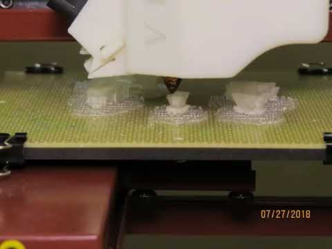
This is a timelapse video of 3d printing the bones of an Australopithecus afarensis child, known as Selam. The original print was completed for a research project; this is a reprint for the purposes of demonstrating the technique. The physical model is the final result of segmenting data from a CT scan of the fossilized remains and digital recreation of the anatomy.